Home

ok elegantní Zbytečný ramus ascendens Perth Útočiště slabika

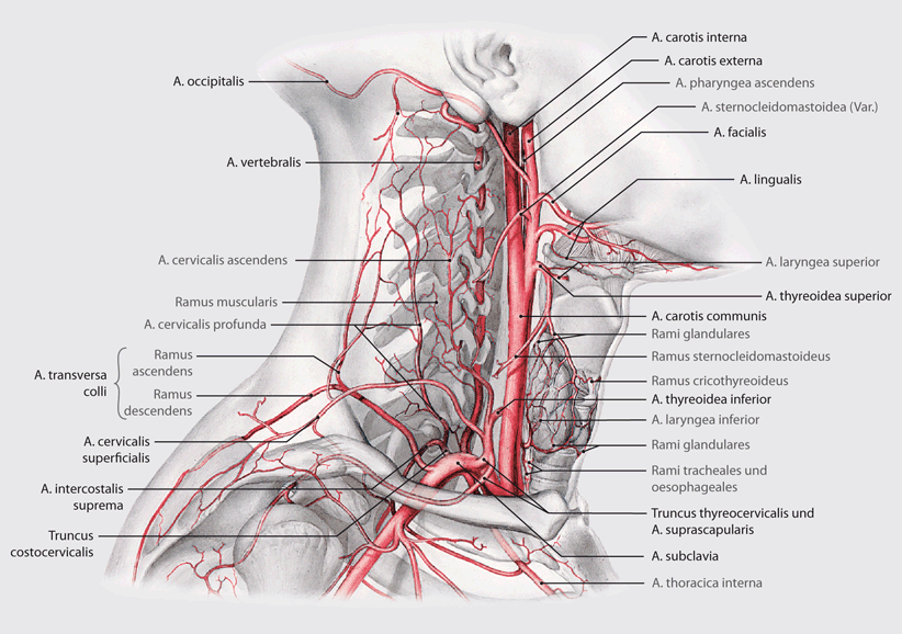
This image is taken from Page 614 of Anatomischer Atlas : … | Flickr

Figure 9 | Gefäßgestieltes Beckenkammtransplantat zur Behandlung der  Femurkopfnekrose | SpringerLink
Figure 9 | Gefäßgestieltes Beckenkammtransplantat zur Behandlung der Femurkopfnekrose | SpringerLink

Figure 56 from Anatomy of the central nervous system. Multiple-choice  questions ("Анатомия центральной нервной системы. Тесты") | Semantic Scholar
Figure 56 from Anatomy of the central nervous system. Multiple-choice questions ("Анатомия центральной нервной системы. Тесты") | Semantic Scholar

Gender-Specific Left–Right Asymmetries in Human Visual Cortex | Journal of  Neuroscience
Gender-Specific Left–Right Asymmetries in Human Visual Cortex | Journal of Neuroscience

Anatomi panosundaki Pin

File:Arterien des Oberschenkels (Schema ventral).svg - Wikimedia Commons

Animals | Free Full-Text | The Heart of the Killer Whale: Description of a  Plastinated Specimen and Review of the Available Literature
Animals | Free Full-Text | The Heart of the Killer Whale: Description of a Plastinated Specimen and Review of the Available Literature

A manual of anatomy . thyreocervicalis) is only a fewmillimeters in length  and divides into three branches: (i) The inferiorthyreoid artery, that  gives branches to the thyreoid gland, muscles ofthe neighborhood, esophagus,
A manual of anatomy . thyreocervicalis) is only a fewmillimeters in length and divides into three branches: (i) The inferiorthyreoid artery, that gives branches to the thyreoid gland, muscles ofthe neighborhood, esophagus,

On the Cranial Osteology of the Lagomorpha

The isolated mandibular ramus - a hitherto rarely described anomaly of the  mandible. Pathogenesis and treatment. | Semantic Scholar
The isolated mandibular ramus - a hitherto rarely described anomaly of the mandible. Pathogenesis and treatment. | Semantic Scholar

Robust optimization of osteosynthesis treatment of mandible fractures under  consideration of inter-individual bone shape variations - PDF Kostenfreier  Download
Robust optimization of osteosynthesis treatment of mandible fractures under consideration of inter-individual bone shape variations - PDF Kostenfreier Download

上行枝 - vet-Anatomy - IMAIOS

Hemispherium cerebri. Fissura longitudinalis cerebri ile serebral  hemisferler birbirinden ayrılırlar. - PDF Free Download
Hemispherium cerebri. Fissura longitudinalis cerebri ile serebral hemisferler birbirinden ayrılırlar. - PDF Free Download

Inferior frontal gyrus - Wikipedia

Definitions of ramus: Synonyms, Antonyms and Pronunciation

3D-Röntgen in der zahnärztlichen Praxis – das Plus an Sicherheit |  Implantologie | DImagazin-aktuell.de
3D-Röntgen in der zahnärztlichen Praxis – das Plus an Sicherheit | Implantologie | DImagazin-aktuell.de
上行枝 - vet-Anatomy - IMAIOS

donor source
donor source

Ascending ramus of the lateral sulcus | Radiology Reference Article |  Radiopaedia.org
Ascending ramus of the lateral sulcus | Radiology Reference Article | Radiopaedia.org

Rami of the Mandible - Dental Technology: How-To, Tips

Arteria femoralis - KenHub Flashcards | Quizlet

Figure 2 | Operative Zugangswege in der rekonstruktiven Gefäßchirurgie der  Arterien | SpringerLink
Figure 2 | Operative Zugangswege in der rekonstruktiven Gefäßchirurgie der Arterien | SpringerLink

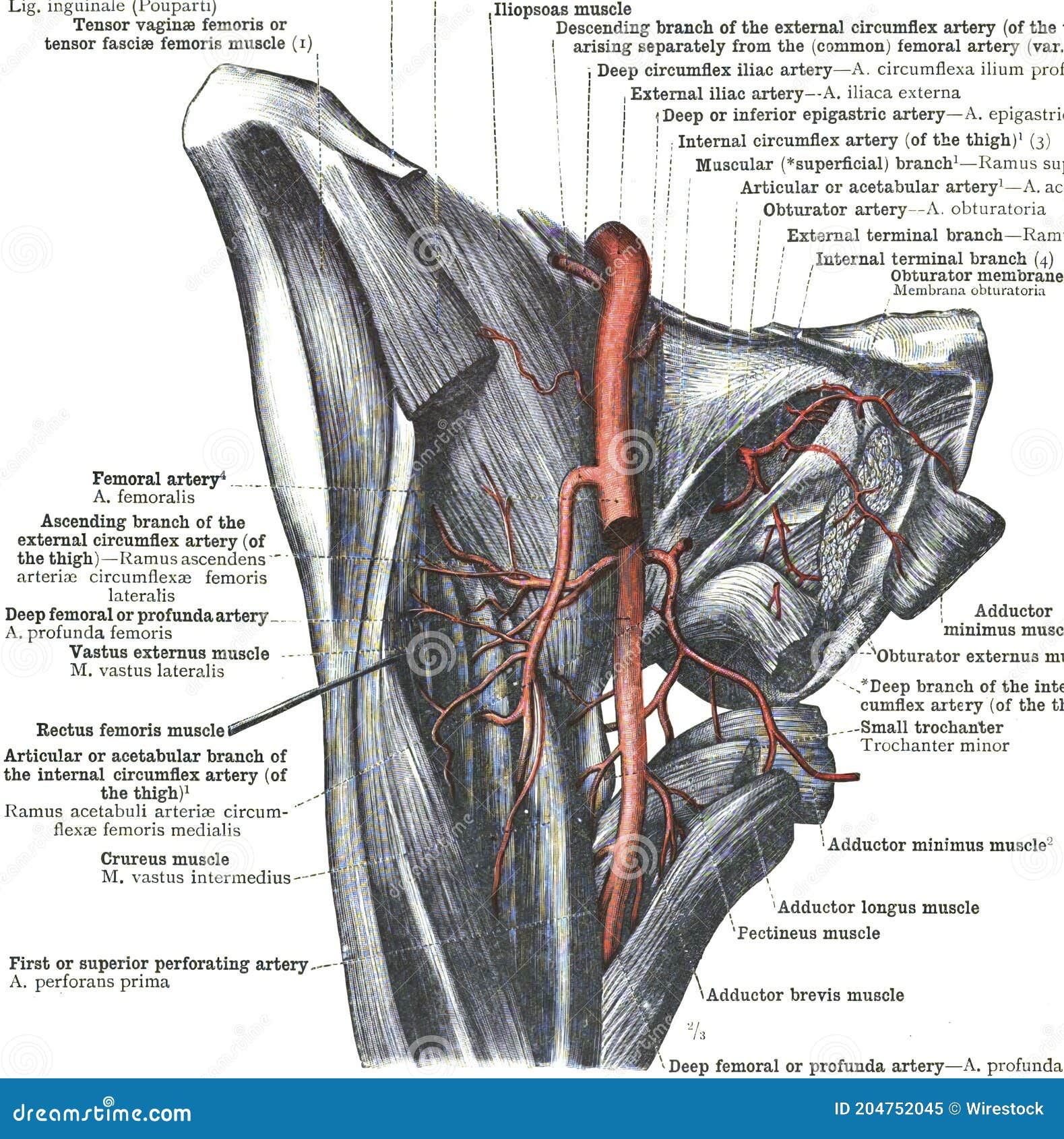
Branches of the Femoral Artery Stock Image - Image of vein, artery:  204752045
Branches of the Femoral Artery Stock Image - Image of vein, artery: 204752045

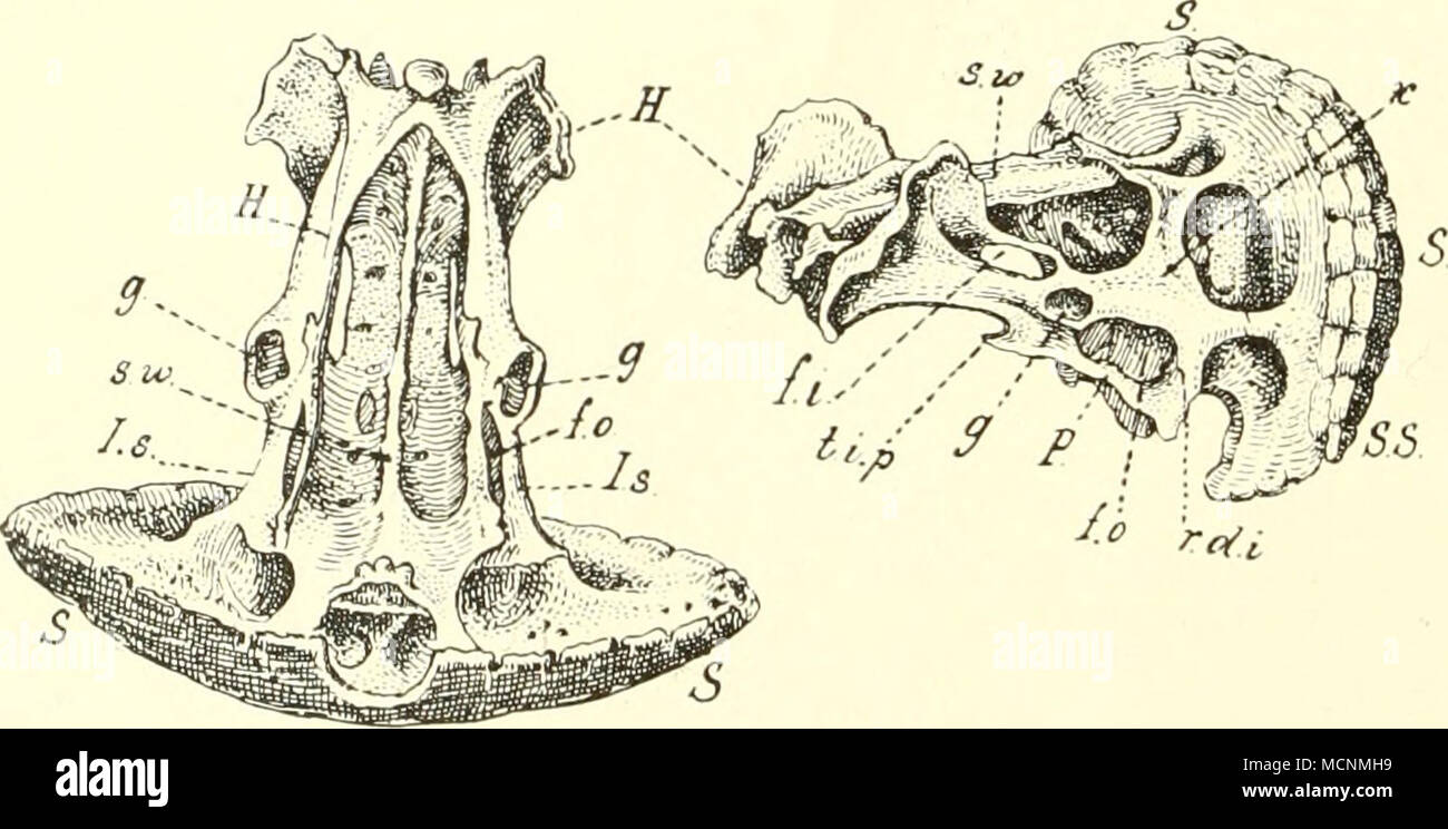
Fig. 336. Becken und Schwanzschild von Chlainydo- phorus truncatus Harl.;  nach Hyrtl, n. Gr.; links von der Ventralfläche, rechts von der rechten  Seite gesehen, fi Fo- ramen ischii; fo Foramen obturatum; ^-
Fig. 336. Becken und Schwanzschild von Chlainydo- phorus truncatus Harl.; nach Hyrtl, n. Gr.; links von der Ventralfläche, rechts von der rechten Seite gesehen, fi Fo- ramen ischii; fo Foramen obturatum; ^-